Seminar Papers
The authors reviewed recent work in the intersection of cognitive science, computational neuroscience and artificial intelligence. Computational models that mimic brain information processing during perceptual, cognitive and control tasks are beginning to be developed and tested with brain and behavioral data.
https://www.nature.com/articles/s41593-018-0210-5
N. Kriegeskorte, P.K. Douglas. Cognitive computational neuroscience. Nat. Neurosci., 21 (2018), pp. 1148-1160
A research team recently found new evidence revising the traditional view of the primate brain’s visual system organization. This remapping of the brain could serve as a future reference for understanding how the highly complex visual system works, and potentially influence the design of artificial neural networks for machine vision. (from the below link)
https://www.sciencedaily.com/releases/2019/08/190821113151.htm
Bing‐Xing Huo, Natalie Zeater, Meng Kuan Lin, Yeonsook S. Takahashi, Mitsutoshi Hanada, Jaimi Nagashima, Brian C. Lee, Junichi Hata, Afsah Zaheer, Ulrike Grünert, Michael I. Miller, Marcello G. P. Rosa, Hideyuki Okano, Paul R. Martin, Partha P. Mitra. Relation of koniocellular layers of dorsal lateral geniculate to inferior pulvinar nuclei in common marmosets. European Journal of Neuroscience, 2019; DOI: 10.1111/ejn.14529
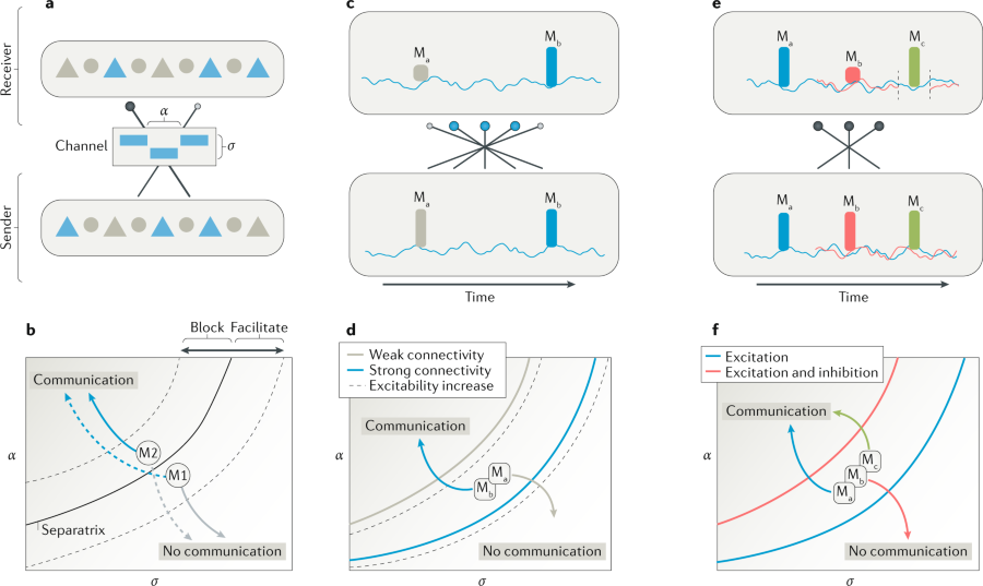
The brain is organized into a network of specialized networks of nerve cells.
For such a brain architecture to function, these specialized networks ―each located in a different brain area― need to be able to communicate with each other. But which conditions are required for communication to take place and which control mechanisms work?
Researchers at the Bernstein Center Freiburg and colleagues in Spain and Sweden are proposing a new model that combines three seemingly different explanatory models. Their conclusions have now been published in Nature Reviews Neuroscience.
https://www.sciencedaily.com/releases/2018/12/181217120046.htm
Researchers have shown that astrocytes ― long-overlooked supportive cells in the brain ― help to enable the brain’s plasticity, a new role for astrocytes that was not previously known.
The findings could point to ways to restore connections that have been lost due to aging or trauma.
https://www.sciencedaily.com/releases/2018/10/181018141035.htm
A popular artificial-intelligence method provides a powerful tool for surveying and classifying biological data. But for the uninitiated, the technology poses significant difficulties.
- Thoughts and discussion about the brain from Peter Bandettini and Eric Wong
In this blog, the professor Eric Wong discussed about “How can we find out how the brain works?”, which is asked from the Cognitive Computational Neuroscience (CCNeuro) conference.
In short, he mentioned that the most typical conceptual approach to understanding the brain is considering the brain is modular. Thus, the modularity is important for understanding the brain from complexity.
The more details is shown in the brain blog (www.thebrainblog.org).
Beside this issues, you could find many neuroscience related thought and discussion from Peter Bandettini and Eric Wong in this blog.