Seminar Papers

Astrocytes help transition the brain from a highly plastic state to one that is more stable.
Separating vascular cell data based on sex helps researchers make new discoveries about why males and females are affected by neurodegenerative* diseases differently. Findings point to differences in the blood-brain barrier between males and females.
*neurodegenerative : the progressive loss of structure or function of neurons, including their death
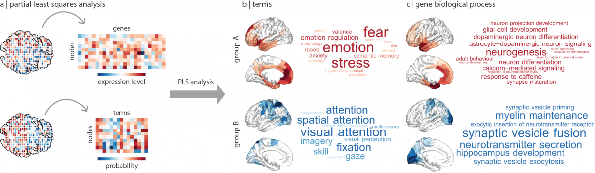
A new study, that utilized machine learning tools, provides a new map that links genetic signatures to functions across the human brain!
Interestingly, they found a clear genetic signal that separated cognitive processes, like attention, from more affective processes, like fear. This separation can be traced to gene expression in specific cell types and molecular pathways, offering key insights for future research into psychiatric disorders.
Cognition, for example, was linked more to the gene signatures of inhibitory or excitatory neurons. Affective processes, however, were linked to support cells such as microglia and astrocytes, supporting a theory that inflammation of these cells is a risk factor in mental illness. The genetic signature related to affect was centred on a brain region called the anterior cingulate cortex, which has been shown to be vulnerable in mental illness.
See the https://www.nature.com/articles/s41562-021-01082-z
Published in the journal Nature Human Behaviour on March 25, 2021
(this study draws a direct link between gene expression and higher brain function, by mapping gene signatures to functional processes across the human brain.)
Johnson, D. D., & Fowler, J. H. (2011). The evolution of overconfidence. Nature, 477(7364), 317-320.

Interestingly, the authors found that overconfidence maximizes individual fitness and populations tend to become overconfident.

The human brain has limited capacity in terms of the data it is able to process and save in its memory. Researchers sought to answer the following question: is the visual system capable of automatic object categorization (i.e., without attention)? To this end, they tested whether the rapid categorical parsing is automatic or requires attention. They found that spatially intermixed objects are parsed into distinct categories automatically.
How Our Visual System Avoids Overloading

Using generative networks and functional magnetic resonance imaging (fMRI), researchers found that brain uses similar visual areas for mental imagery and vision, but it uses low-level visual areas less precisely with mental imagery than with vision. Namely, there are distinct codes for seen and mental images in the brain which can be captured by generative networks.
Is What I See, What I Imagine? The Neural Overlap Between Vision and Imagination
Breedlove, J. L., St-Yves, G., Olman, C. A., & Naselaris, T. (2020). Generative Feedback Explains Distinct Brain Activity Codes for Seen and Mental Images. Current Biology.

The team found that those with training in mindfulness-based attention and training (MBAT) were more successful in controlling the BCI, both initially and over time. Interestingly, the researchers found that differences in brain activity between the two sample groups corresponded directly with their success. The meditation group showed significantly enhanced capability of modulating their alpha rhythm, the activity pattern monitored by the BCI system to mentally control the movement of a computer cursor.
His findings are very important for the process of BCI training and the overall feasibility of non-invasive BCI control via EEG. This work shows that just a short period of MBAT training can significantly improve a subject’s skill with a BCI. This suggests that education in MBAT could provide a significant addition to BCI training.
https://neurosciencenews.com/meditation-mind-control-17071/
Mindfulness Improves Brain–Computer Interface Performance by Increasing Control Over Neural Activity in the Alpha Band James R Stieger, Stephen Engel, Haiteng Jiang, Christopher C Cline, Mary Jo Kreitzer, Bin He

Researchers have uncovered the neural mechanism underlying rumination. The study reports when rumination occurs, coupling between the core and medial temporal lobe subsystems of the default mode network becomes elevated while coupling between the core and dorsomedial prefrontal cortex decreases. According to this study, they computed the Pearson’s correlation during the activity among different brain regions. Results revealed that couplings between the core and the medial temporal lobe (MTL) subsystems of the default mode network (DMN) were elevated while the other couplings were decreased.

Researchers conducted a first-of-its-kind study designed to investigate brain connectivity in 130 mammalian species. The intriguing results, contradicting widespread conjectures, revealed that brain connectivity levels are equal in all mammals, including humans.
Their study revealed a universal law: Conservation of Brain Connectivity which denotes that the efficiency of information transfer in the brain’s neural network is equal in all mammals, including humans. They also discovered a compensation mechanism which balances the connectivity in every mammalian brain. This mechanism ensures that high connectivity in a specific area of the brain, possibly manifested through some special talent (e.g. sports or music) is always countered by relatively low connectivity in another part of the brain.
In future projects they will investigate how the brain compensates for the enhanced connectivity associated with specific capabilities and learning processe.
https://www.sciencedaily.com/releases/2020/07/200720112216.htm
Yaniv Assaf, Arieli Bouznach, Omri Zomet, Assaf Marom, Yossi Yovel. Conservation of brain connectivity and wiring across the mammalian class. Nature Neuroscience, 2020; 23 (7): 805